A clock position, or clock bearing, is the direction of an object observed from a vehicle, typically a vessel or an aircraft, relative to the orientation of the vehicle to the observer The vehicle must be considered to have a front, a back, a left side and a right side These quarters may have specialized names, such as bow and stern for a vessel, or nose and tail for an aircraftCase Discussion MACROSCOPIC DESCRIPTION "Left breast 1 o'clock, 7cm FN" Four cores 30mm in total length A1 MICROSCOPIC DESCRIPTION Sections show multiple cores of breast parenchyma which shows an invasive carcinoma with morphological features in keeping with a BRE grade 2 tumor DIAGNOSIS Left breast 1 o'clock 7cm FN Invasive carcinoma NSTBreast cancer mortality Women with invasive cancers of 1 cm or smaller have a 95% chance of survival at 10 years, while those with invasive cancers 1–2 cm and 2–5 cm in size have, respectively, 85% and 60% survival at 10 years (1) For large invasive cancers, survival is dependent on tumor grade (1) However, survival is high for women

Individualized Treatment Analysis Of Breast Cancer With Chronic Renal Ott
Where is 9 o'clock left breast
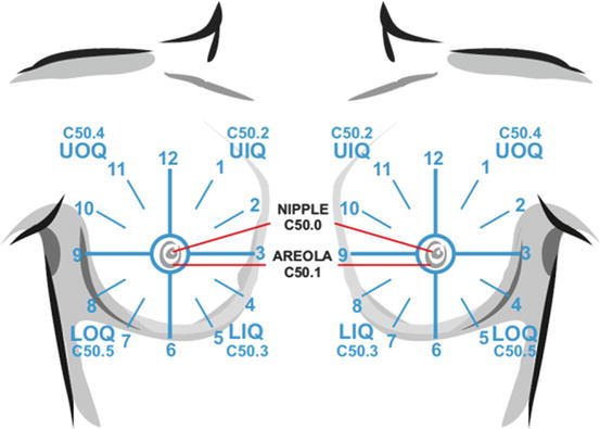
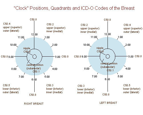
Where is 9 o'clock left breast-Code C509 (Breast, NOS) in this situation Code the primary site to C508 when O'Clock Positions and Codes Quadrants of Breasts 2 11 12 1 1 10 9 8 7 7 6 5 4 3 2 11 12 10 6 5 3 RIGHT BREAST LEF T BREAST UOQ UIQ UIQ UOQ LOQ LIQ LIQ LOQ C504In contrast to breast cancer tumors, benign lumps are often squishy or feel like a soft rubber ball with welldefined margins They're often easy to move around (mobile) and may be tender 4 Breast infections can cause redness and swelling Sometimes it can be difficult to tell the difference between mastitis and inflammatory breast cancer
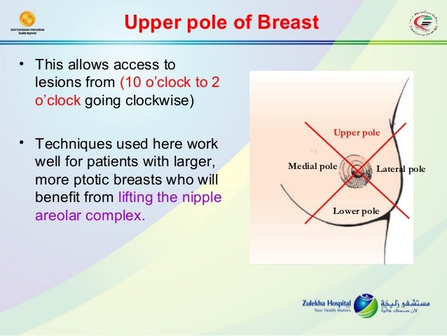


Breast Oncoplastic Surgery
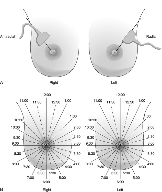
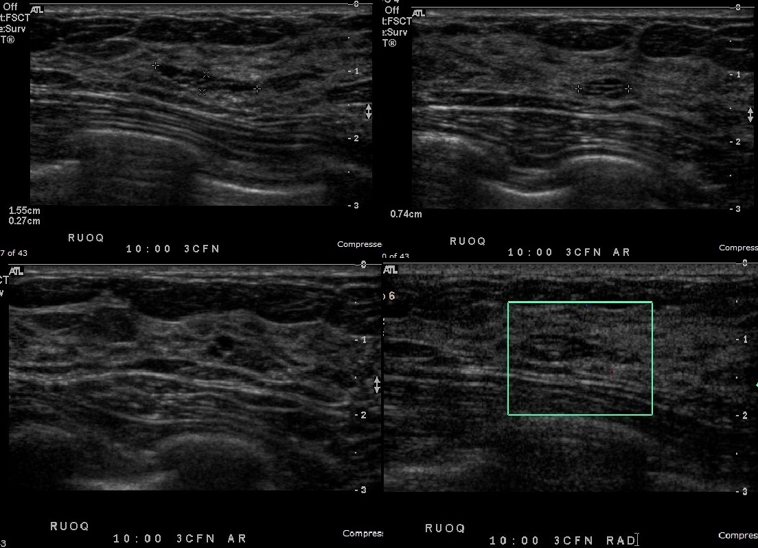
Figure 11 Accurate annotation on a breast US image The description of a lesion should include laterality (right or left breast), the clockface location (estimated as 10 o'clock 1000), the distance from the nipple (stated as N 34cm) and the orientation of the probe (radial Rad, antiradial, transverse, or longitudinal) Figure 11I noticed a pink spot at around the 10 oclock position of my left breast 3 days ago i had had pain in the right breast a few days before i did wear a different bra the 2 days before i saw this spot on the left breast and did a breast check the night before Show More Show LessUnspecified lump in the left breast, upper outer quadrant 18 New Code 19 21 Billable/Specific Code N6321 is a billable/specific ICD10CM code that can be used to indicate a diagnosis for reimbursement purposes;
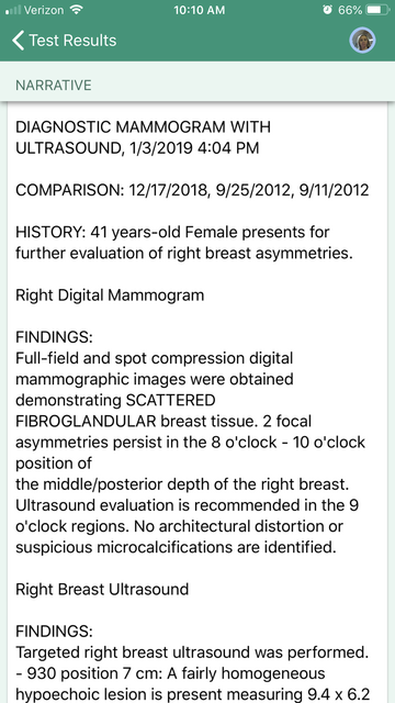
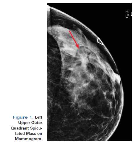
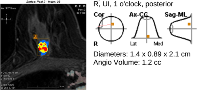
Unspecified lump in the right breast, upper inner quadrant 18 New Code 19 21 Billable/Specific Code N6312 is a billable/specific ICD10CM code that can be used to indicate a diagnosis for reimbursement purposes;What does Scattered Fibroglandular Densities Mean?Standard twoview mammography followed by additional mammographic projections is an effective way to demonstrate the spiculated mass and to classify the prepectoral lesion as category BIRADS 4 or 5 Additional ultrasonography can overcome the mimicry of invasive lobular breast carcinoma at mammogra
The 10o'clock position on a mammogram and sees a similar lesion at the 8o'clock position on ultrasound, they are very likely to be different lesions unless the mammographic posi tioning was initially incorrect or the findings were misinterpreted 2–4 Triangulation does have its weaknesses, mainly because of the positioning of the 90° mediolateral view compared with the positionLisa Jacobs, MD, Johns Hopkins breast cancer surgeon, and Eniola Oluyemi, MD, Johns Hopkins Community Breast Imaging radiologist, receive many questions about how to interpret common findings on a mammogram reportThe intent of the report is a communication between the doctor who interprets your mammogram and your primary care doctor However, this report is oftenGiven this guidance, a 12 o'clock right breast mass can be reported as ICD10 code N6315, Unspecified lump in right breast, overlapping quadrants, or as dual ICD10 codes for overlapping quadrants, N6311, Unspecified lump in the right breast, upper outer quadrant, and N6312, Unspecified lump in the right breast, upper inner quadrant


The Role Of Ultrasound In Breast Imaging Document Gale Onefile Health And Medicine


File Breast Clock Positions Svg Wikimedia Commons
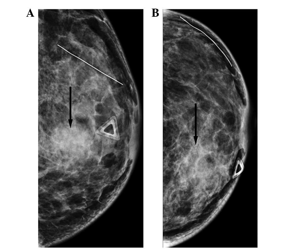
• The hardest part of the breast to image (and the area most often missed on the MLO) is the posterior medial breast If done properly (offsetting the IR into the contralateral breast) you will be able to get deeper against the chest wall • There is no issue of the contralateral breast impeding the path of the compression paddleThe imaging of the right breast is normal ( not shown ) The left breast demonstrates a 1cm isodense oval mass in the posterior depth of the left breast at the 6 to 7 o'clock location, 10 cm from the nipple ( Fig 708, Fig 709, Fig 7010 ) A fatty hilum is suggested on the lateromedial (LM) spotcompression digital breast tomosynthesisOne lump was aspirated (upper inner right breast marked by arrows in A and B), yielding malignant cytology (D) Repeat ultrasonography of the known malignant mass in the right breast at 2o'clock position shows it to be indistinctly marginated and lobulated both without and (E) with spatial compounding Any lesion documented on
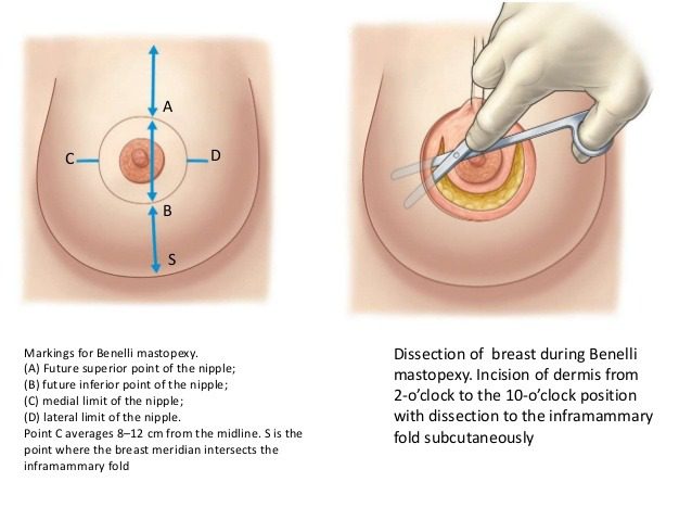


Advances In Breast Lifts Orlando Florida Best Plastic Surgeon
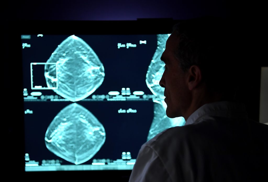


Breast Cancer Signs Symptoms And Understanding An Imaging Report Saint John S Cancer Institute
Breast ultrasound allows its detection and the correct diagnosis of cyst has been reported to be almost 100% 5% of breast ultrasound examinations report complex cyst 7 Complex breast cysts have a malignancy rate of 03% among breast neoplasms, it still has had a substantial probability of being malignant (23% and 31% in 2 series) 7 PathologyTumor processes, which include breast carcinoma, often begin their development in the upper outer quadrant (in 45% of cases) Relatively rare diseases of the gland are found in the upper inner quadrant (up to 15%), in the lower outer quadrant (up to 10Breast cancer, ultrasonography The patient in Images 2628 also had a 7mmdiameter cyst at the 10o'clock position in the right breast (black central structure) Note the wellcircumscribed
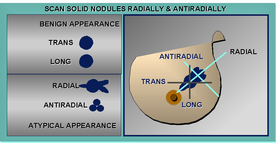


Sonography Of The Breast


Right Breast 1 O Clock Position 2 Cm From Nipple Deep To The Skin Download Scientific Diagram
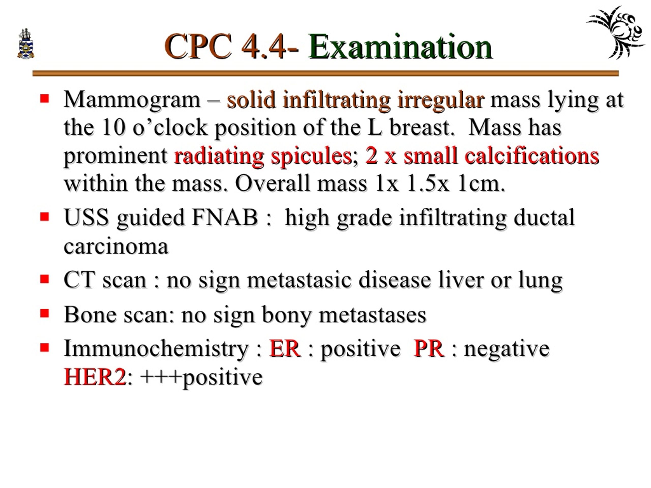
10 o'clock position on mammogram () Recent clinical studies Diagnosis Cavernous hemangioma of the breast mammographic and sonographic findings and followup in a patient receiving hormonereplacement therapy Mesurolle B, Wexler M, Halwani F, Aldis A, Veksler A, Kao E J Clin Ultrasound 03 Oct;31(8)4306 doi /jcuI'm 53 had breast augmentation 17 years ago During my yearly mammogram in May 17 nodules were detected as well as a small cyst in the left 3'oclock measuring 45mm I was told to have routine mammographic followup Told Discovery rules changedBreast cancer, ultrasonography Mediolateral oblique digital mammogram of the right breast in a 66yearold woman with a new, opaque, irregular mass approximately 1 cm in diameter The mass has spiculated margins in the middle third of the right breast at the 10o'clock position


Www Ijsurgery Com Index Php Isj Article Viewfile 4785 3094
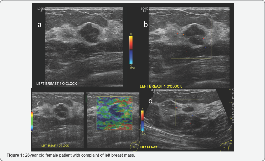


Cancer Therapy And Oncology International Journal Ctoij
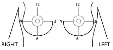
3 o'clock position (right breast) is towards the middle of your chest3 o'clock position (left breast ) is towards your armpitHope I've been helpfulRegards Comment japdip It's Clockwise in both breasts Comment nannotnancy Thank you so much!(b) Bilateral craniocaudal mammograms reveal a focal asymmetric density at the 12 o'clock position in the right breast (arrow) Additional mammography and US were performed due to suspected occult malignancyMy tumour was located at the 12o'clock position in my right breast Given the size and solid shape of the mass, my surgeon performed a lumpectomy The breast cancer surgery process often includes a sentinel lymph node biopsy (SLNB) (FCKING OUCH), where a chain of nodes are removed to test for cancer spread within the lymphatic system


Breast Cancer Topic Quadrants Of Breast Cancer Survey Also


Diagnosis And Management Of Benign Breast Disease Glowm
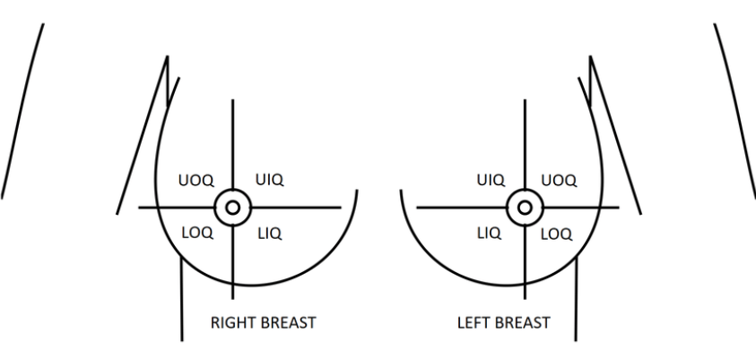
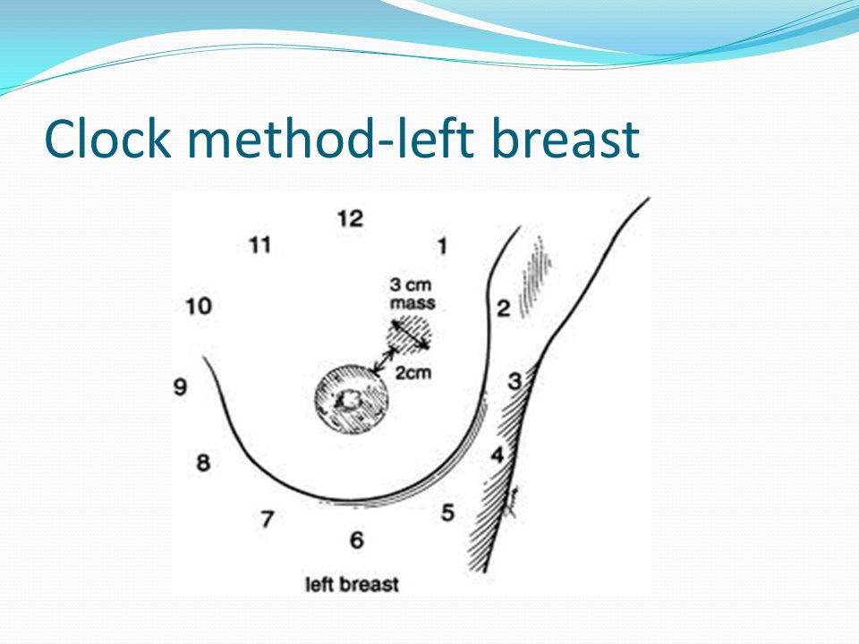
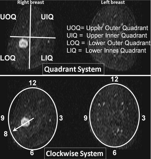
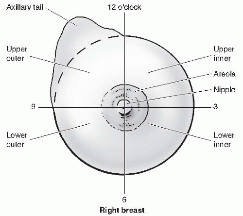
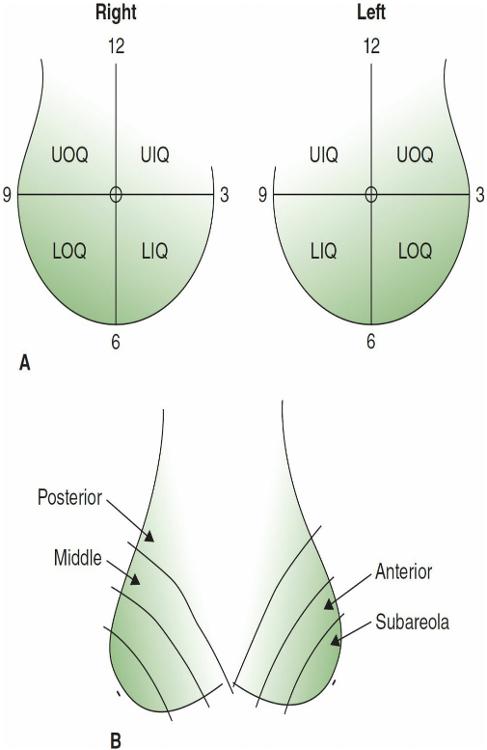
The outer left breast is at 3 o'clock and the outer right breast is at 9 o'clock In the left breast the upper outer quadrant is between 12 and 3 o'clock The radiologist will also describe the size and location of a finding by indicating the distance from the nipple in centimeters Centimeters are smaller than an inchCorresponds to an irregular mass lesion with indistinct margins at the 10 o'clock position, 30 mm from the nipple Combined imaging category 5 Malignant Architectural distortion and stellate lesion Architectural distortion represents lesions seen on mammography that cause distortion of the normal architecture of the breast parenchymaCode C509 (Breast, NOS) in this situation O'Clock Positions and Codes Quadrants of Breasts 2 11 12 1 1 10 9 8 7 6 5 7 4 3 2 11 12 10 6 5 4 3 RIGHT BREAST LEF T BREAST UOQ UIQ UIQ UOQ LOQ LIQ LIQ LOQ C504 C502 C502 C504 C505 C503 C505 C503 C500 C501


Usefulness Of Postoperative Surveillance Mr For Women After Breast Conservation Therapy Focusing On Mr Features Of Early And Late Recurrent Breast Cancer


Nucleus Iaea Org Hhw Radiology Clinical Applications Specialities And Organ Systems Breast Imaging Guidelines And Literature Suggested Articles Reading A Mammogram Pdf
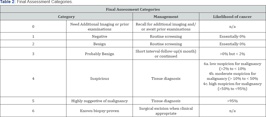
This is the American ICD10CM version of N6312 other international versions of ICD10Of diagnostic mammography and breast ultrasonography in detection and differentiation of posterior breast cancers Methods The study included 40 women with palpable, histopathological confirmed posterior breast cancer Mammographic and ultrasonographic features were defined according to Breast Imaging Reporting and Data System (BIRADS) lexiconA lesion located in the upper outer quadrant of the right breast is located in the 1 5o'clock position 2 2o'clock position 3 10o'clock position 4 7oc'clock position


Spie Org Samples Pm211 Pdf


Contrast Enhanced Dedicated Breast Ct Detection Of Invasive Breast Cancer Preceding Mammographic Diagnosis Sciencedirect
Abstract BACKGROUND Metaplastic carcinoma is a rare form of breast carcinoma that often is confused with other benign and malignant entities TheThe 21 edition of ICD10CM N6312 became effective on ;Ie Notice how UIQ is either 12 to 3


Diagnostic Breast Imaging Clinical Breast Imaging A Patient Focused Teaching File 1st Edition
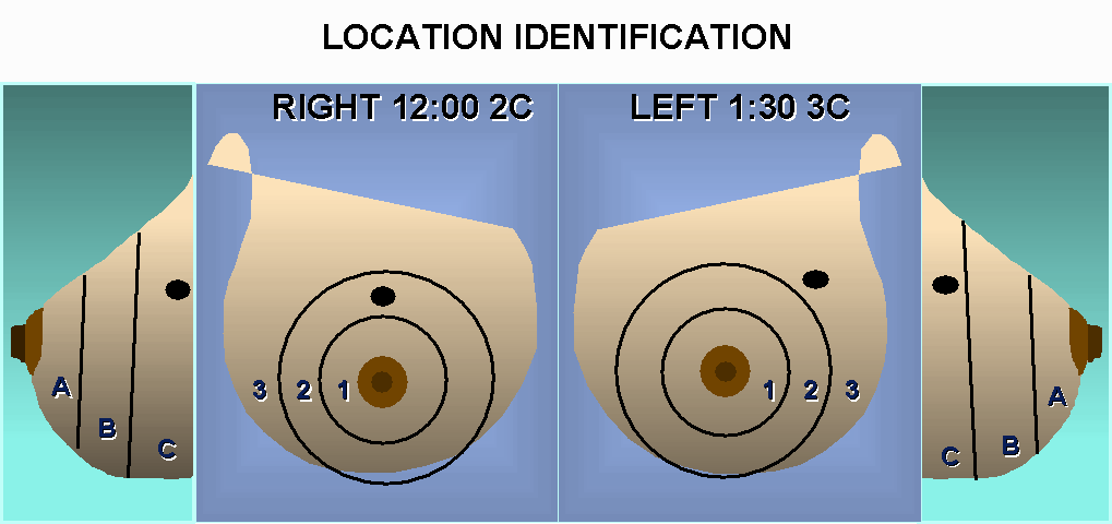


Sonography Of The Breast
In the Eleventh Edition Education slides Neoplasms, there is a diagram with clock positions with an example of documentation of a breast lesion at 6 o'clock coded to C508 Overlapping lesion of breastThe breast on the mammogram The mammogram read as negative The patient was offered an ABUS exam due to her heterogeneously dense breast tissue Investigation and Diagnosis ABUS detected a right breast irregular hypoechoic mass with angular margins and posterior acoustic shadowing at the 1030 o'clock position, measuring 15 x 16 x 11 cm, 7 cmBreasts are made out of both fatty tissue and fibroglandular tissue (see image) The level of thickness is controlled by the mammogram The more fibroglandular tissue, the denser the breast Fatty tissue is fundamentally dark on the mammogram and fibroglandular tissue is white Breasts are isolated into 4 classifications
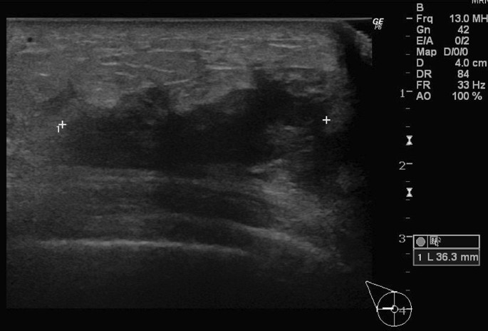


Inflammatory Breast Cancer Associated With Amyopathic Dermatomyositis A Case Report Surgical Case Reports Full Text


Breast Coding Guidelines Surveillance Epidemiology And Flip Ebook Pages 1 4 Anyflip Anyflip
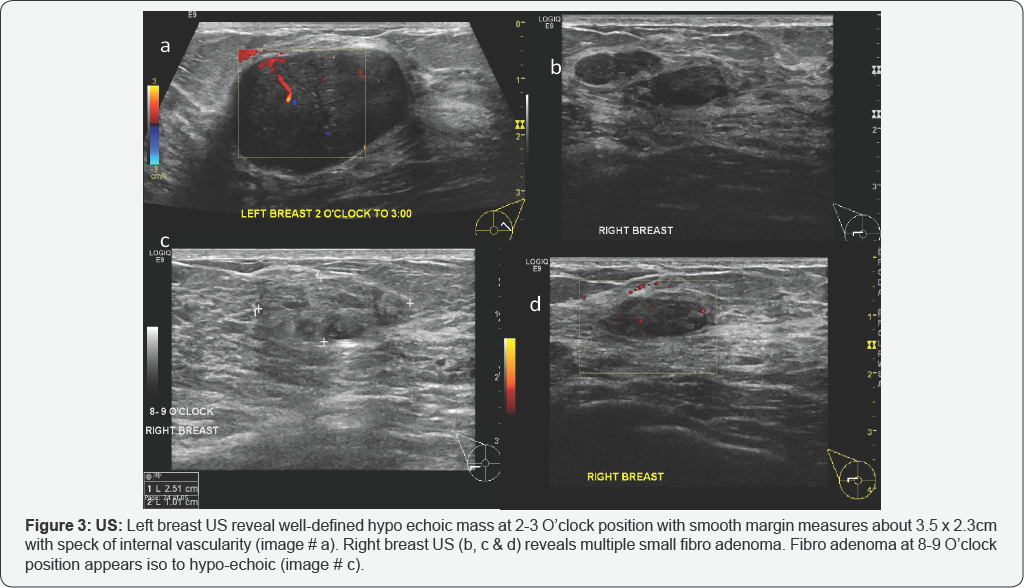
In the Introduction, Dr Ellis wrote, "Due to these positional differences, a lesion reported at the 10o'clock position of the breast with mammography may be much closer to the 8o'clock position with sonography" Although confusion between different quadrants may occur for the lesions near the 3, 6, 9, or 12o'clock positions, the 10o'clock and 8o'clock positions are in completelyStandard lumpectomy in the lower breast frequently results in significant defect and downpointing of the nipple For radially oriented lesions in the lower hemisphere of the breast (4o'clock to 8o'clock position, going clockwise), reduction mastopexy lumpectomy is a useful technique for excision that prevents deviation of nipple (Fig 147)Mammogram Circumscribed mass in the lower inner left breast which persists on spot compression views (not shown) Ultrasound Echogenic solid mass with marked vascular flow on color Doppler imaging at 8 o'clock 10 cm from the nipple measuring 6 x 4 x 6 mm, corresponding to the site of the mammographic mass


Chapter 3 Lange Improved Flashcards Quizlet


Q Tbn And9gcth39qrs02wrthtpk Uisqk0p0rnefiz Yi0mmwn2cv4eblwxg Usqp Cau
There is on benign appearing solid lesion at 10 o'clock position in the (R) breast measuring from the nipple This is stable since the previous study In the (L) breast, there is a solid lesion at 4 o'clock position, 5 cm from the nipple It has microlobulated margins andNan Comment Next QuestionWhen the breast is lifted up to position it on the image receptor, the tissue along the periphery of the breast should be palpated and additional special views of any thickening or mass that would not be imaged on the routine twoview mammogram should be obtained 30° Superolateralto


Breast Oncoplastic Surgery
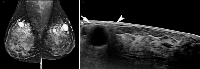


Cleopatra S Cosmetics A Case Of Silicone Breast Injection Granulomata
Breast asymmetry refers to when one breast is a different size or shape than the other A mammogram or breast cancer screening may show asymmetrical breast size or densityThis is the American ICD10CM version of N6321 other international versions of ICD10The 21 edition of ICD10CM N6321 became effective on ;


Quantitative Analysis Of Enhancement Intensity And Patterns On Contrast Enhanced Spectral Mammography Scientific Reports


Breast Cancer Topic Interpreting Your Report
On physical exam, a 10mm mass was palpated in the left breast at the 10 o'clock position, medial to the areola A screening mammogram performed four months earlier demonstrated scattered fibroglandular densities bilaterally with mild stable architectural distortion in the right retroareolar region at the site of prior benign lumpectomy, and bilateral benign secretory calcifications (Figs•Right Breast UIQ –12 to 3 LIQ –3 to 6 LOQ – 6 to 9 UOQ –9 to 12 •Left Breast UIQ –9 to 12 UOQ –12 to 3 LOQ –3 to 6 LIQ –6 to 9 R v L matters!In the left breast, the nerve generally exits the chest wall at the lateral border of the pectoralis minor, enters the posteriorlateral surface of breast at approximately the 4 o'clock position, and then traverses the glandular tissue to the inner areola along the 5 o'clock axis In the right breast, the nerve generally enters the


National Program Of Cancer Registries Education And Training Series How To Collect High Quality Cancer Surveillance Data Ppt Download


Breast Metastasis From Nasopharyngeal Carcinoma A Case Report And Review Of The Literature
I would recommend to use 2 diagnoses the quadrants that the mass/lump spans ie right side 12oclock would be N6311 for upper outer AND N6312 for upper inner It's not unspecified I am using the logic that if a patient had a left ovarian cyst and a right ovarian cyst, you would code both, not unspecifiedFig 61 At the 12 o'clock position of the left breast and the 3 o'clock position of the right breast, there are two oval masses (arrows) The spot compression views demonstrate that the margins of the right mass are well defined, and the margins of the left mass are ill defined (A) Right MLO mammogram (B) Left MLO mammogramTwenty patients demonstrated a change in asymmetric tissue size, most commonly in the upper outer quadrant, followed by the axillary tail, the 12 o'clock position and the inner part of the breast


Http Static pc Com A3c7c3fe 6fa1 4d67 8534 A3c9c15fa0 C7b39f96 0935 4e94 84b8 C4a6d9d86abd 3739cb 8b1c 4e15 6b E6192eec5078 Pdf


Icd 10 Cm Coding Overlapping Breast Quadrants Coffee With A Medical Coding Auditor
Tumor size is an important factor in breast cancer staging, and it can affect a person's treatment options and outlook Tumors are likely to be smaller when doctors detect them early, which canSimply refers to the location approximately midway between the nipple and the chest wall on the mammogram An asymmetry is a potential abnormalty You probably need extra mammo views and maybe an ultrasound to sort it out Most of these turn out to be nothing, but you need to complete the evaluation to be sure


Ultrasonography At The 12 O Clock Position Of The Left Breast Revealed Download Scientific Diagram


Coding Breast Mass Becomes More Specific For Coders


A 55 Year Old Woman With New Triple Negative Breast Mass Less Than 2 Cm On Both Mammogram And Ultrasound


Cancer Therapy And Oncology International Journal Ctoij


Healthy Breast With Ultrasound Radiology Key


Diagnostics Free Full Text A Proposed Dedicated Breast Pet Lexicon Standardization Of Description And Reporting Of Radiotracer Uptake In The Breast Html


Fcds Med Miami Edu Downloads Naaccr Webinars Breast Breast final Pdf


Diabetic Mastopathy In An Elderly Woman Misdiagnosed As Breast Cancer A Case Report And Review Of The Literature


Preoperative Tumor Size Measurement In Breast Cancer Patients Which Threshold Is Appropriate On Computer Aided Detection For Breast Mri Cancer Imaging Full Text
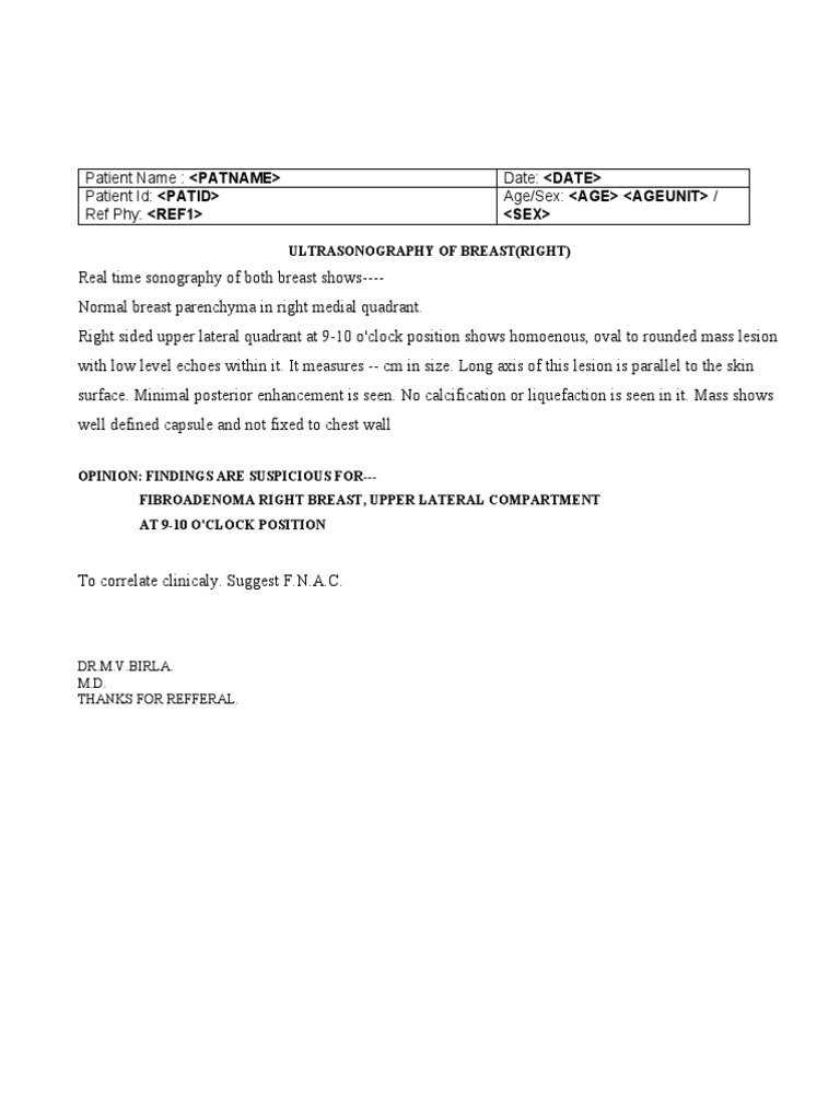


Ultrasonography Breast Single Fibroadenoma
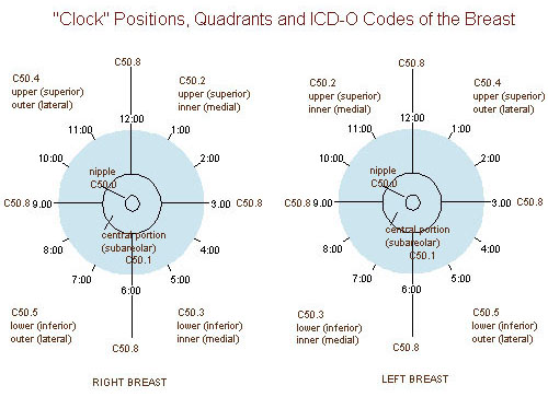


Seer Training Quadrants Of The Breast


Clock Position Wikipedia


2


Seer Cancer Gov Manuals 21 Appendixc Coding Guidelines Breast 21 Pdf
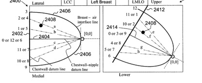


Gallery Formats Parvizbagloo


1


Coding Breast Mass Becomes More Specific For Coders


Antr510 027 Anatomy Of The Breast Msu Mediaspace


Molecular Breast Imaging In Breast Cancer Screening And Problem Solving Radiographics
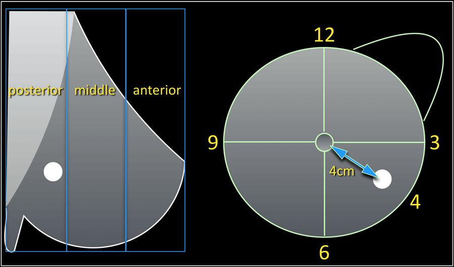


The Radiology Assistant Bi Rads For Mammography And Ultrasound 13


Ieeexplore Ieee Org Iel7 Pdf


O Clock Position Of A Lesion On A Right Breast As Identified By Download Scientific Diagram


Lactational Breast Changes Lobular Hyperplasia Mimicking Masses How Can We Differentiate From True Pathological Masses


Homeoconsult Rd Breast Lump Tumor Or Fibroadenoma Cure With Homeopathy


Ppt Pathology Of Breast Disorders Powerpoint Presentation Free Download Id


Individualized Treatment Analysis Of Breast Cancer With Chronic Renal Ott
/know-your-breast-tumor-size-4114640-FINAL-f17fb19bf9214d20937d07bd41524ac7.png)


Breast Tumor Size And Staging


Breast Cancer Signs Symptoms And Understanding An Imaging Report Saint John S Cancer Institute


Figure 9 Use Of Bismuth Shield For Protection Of Superficial Radiosensitive Organs In Patients Undergoing Computed Tomography A Literature Review And Meta Analysis Springerlink


Osa Three Dimensional In Vivo Fluorescence Diffuse Optical Tomography Of Breast Cancer In Humans


Identification Of A Brca2 Mutation In A Turkish Family With Early Onset Breast Cancer Celik 18 Clinical Case Reports Wiley Online Library


Cancer Therapy And Oncology International Journal Ctoij


Breast Ultrasonography Practice Essentials Technique Invasive Ultrasonography


26 Year Old With Palpable Mass Near The 12 00 O Clock Position Of The Download Scientific Diagram


Www Alabamapublichealth Gov Ascr Assets 17issue1 Pdf


Figure 2 From Efficacy Of Pectoral Nerve Block Type Ii For Breast Conserving Surgery And Sentinel Lymph Node Biopsy A Prospective Randomized Controlled Study Semantic Scholar


Missed Breast Cancer Effects Of Subconscious Bias And Lesion Characteristics Radiographics


Arrs Breast Case Of The Week


1


Journal Of Lancaster General Health Depart3 V1i2


Breast Mass Radiology Key


Electrical Impedance Spectroscopy For Breast Cancer Diagnosis Clinical Study


Breast 34 Fibrocystic Diseasechange Pathology Harmone Induced Acinar


Coding Breast Mass Becomes More Specific For Coders


Imaging Findings For Malignancy Mimicking Nodular Fasciitis Of The Breast And A Review Of Previous Imaging Studies Topic Of Research Paper In Clinical Medicine Download Scholarly Article Pdf And Read For Free


Determining The Thermal Characteristics Of Breast Cancer Based On High Resolution Infrared Imaging 3d Breast Scans And Magnetic Resonance Imaging Scientific Reports


Ultrasound Of The Breast Part 1 Ppt Video Online Download


Invasive Micropapillary Carcinoma Of The Breast Radiology Case Radiopaedia Org


Lange Q A Chapter 3 Anatomy Physiology And Pathology Of The Breast Flashcards Quizlet


Www Bjcancer Org Sites Oldfiles Library Userfiles Pdf 137 Pdf


Secretory Breast Carcinoma A Report Of Three Cases And A Review Of The Literature


Www Biobran Org Uploads Case Study 3a1d32a1d152e7f1b1c5818d322b81ac Pdf


Fcds Med Miami Edu Downloads Naaccr Webinars Breast Breast final Pdf


2


Antr510 027 Anatomy Of The Breast Msu Mediaspace


Cureus Metastatic Malignant Melanoma With Occult Primary Presenting As Breast Mass A Case Report And Literature Review


Clock Position Wikipedia


Breast Mri For Diagnosis And Staging Of Breast Cancer Springerlink


Www Birpublications Org Doi Pdf 10 1259 Bjr


Epos Trade


Breast Cancer Topic If You Are Waiting Please Let Me To Share My Experience


Diagnostic Breast Imaging Clinical Breast Imaging A Patient Focused Teaching File 1st Edition


Factors Affecting Visualization Rates Of Internal Mammary Sentinel Nodes During Lymphoscintigraphy Journal Of Nuclear Medicine


How To Self Examine Your Breast You Me Medicine


Breast Diseases Obgyn Key


Spie Org Samples Pm211 Pdf


Nonvisualization Of Sentinel Node By Lymphoscintigraphy In Advanced Breast Cancer Topic Of Research Paper In Clinical Medicine Download Scholarly Article Pdf And Read For Free On Cyberleninka Open Science Hub


Experts To Consider All Aspects Of Breast Ultrasound European Society Of Radiology


Methylene Blue Dye Related Changes In The Breast After Sentinel Lymph Node Localization Kang 11 Journal Of Ultrasound In Medicine Wiley Online Library


1


Print Anatomy Physiology And Pathology Of The Breast Mammography Flashcards Easy Notecards


Syngo Breast Care


Recent Comprehensive Review On The Role Of Ultrasound In Breast Cancer Management Leaders In Pharmaceutical Business Intelligence Lpbi Group


Automated Whole Breast Segmentation For Hand Held Ultrasound With Position Information Application To Breast Density Estimation Sciencedirect


Epos Trade


Breast Mri Identified Lesions During Neoadjuvant Therapy Are Largely Benign


